Practice Areas/ Case Types
Dr. Milosavljevic provides radiology expert review services tailored to the needs of each client, including matters involving breast imaging, stroke and other neuroradiology findings, fractures and traumatic injury, missed or delayed diagnosis, emergency imaging findings, communication of critical results, follow-up recommendations, and standard-of-care issues. If your matter involves a different type of imaging issue, she would be pleased to discuss your specific needs further.
-
Breast cancer on mammography, ultrasound, or MRI
Lung cancer on chest X-ray or CT
Renal, pancreatic, liver, and bowel malignancies on US, CT or MRI
Gynecologic malignancies on US, CT or MRI
Metastatic disease missed on follow-up CT, PET-CT
-
missed or delayed breast cancer diagnosis
BI-RADS categorization
mammography interpretation
breast ultrasound interpretation
breast MRI interpretation
screening vs diagnostic workflow
biopsy recommendation / biopsy delay
imaging-pathology discordance
post-biopsy management
-
intracranial hemorrhage
stroke or large vessel occlusion
cervical spine injury
solid organ injury
fracture/ dislocation
bowel perforation or obstruction
appendicitis
ectopic pregnancy
pulmonary embolism
aortic emergency
-
lung cancer
pulmonary embolism
pneumonia vs mass
pleural process
mediastinal mass
aortic pathology
incidental lung nodule follow-up recommendations
-
ectopic pregnancy
ruptured ectopic pregnancy
ovarian torsion
early pregnancy loss
adnexal/ ovarian mass
placenta accreta or placental abnormality
abnormal endometrial thickening
endometrial cancer
Polycystic ovarian syndrome (PCOS)
-
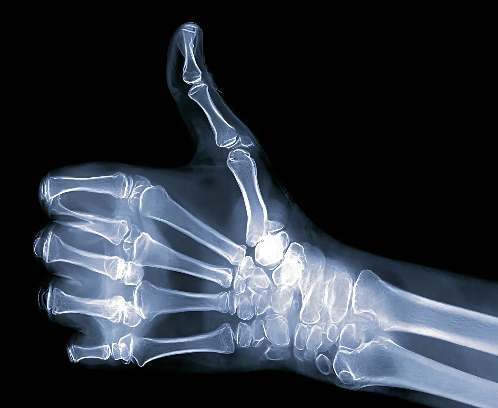
missed fracture or dislocation
occult fracture
osteomyelitis
septic joint
trauma follow-up
-
venous thrombosis
dissection or aneurysm
carotid stenosis grading
-
acute stroke imaging interpretation, including ASPECTS scoring and evaluation of ischemic change
intracranial hemorrhage, including subdural, epidural, and subarachnoid hemorrhage
aneurysms and other intracranial vascular abnormalities
brain tumors and other intracranial mass lesions
delayed diagnosis or missed findings on spine MRI
spinal cord compression
epidural abscess
cauda equina syndrome
-
This includes:
failure to communicate critical findings
inadequate documentation of communication
ambiguous report language
delayed final report
discrepancy between preliminary and final interpretation
failure to contact ordering clinician
inadequate follow-up recommendation
-
incidental lung nodule follow-up
suspicious breast lesion workup
renal/ adrenal/ pancreatic incidentalomas
overdue repeat imaging
missed recommendation for biopsy or advanced imaging
-
contrast reaction management
contrast extravasation
gadolinium-related risk issues
renal-risk screening disputes
radiation exposure / dose concerns
fluoroscopy skin injury
CT radiation counseling / consent issues
-
missed diagnosis
delayed diagnosis
misdiagnosis
false positive interpretation
failure to detect interval change
comparison-study error
undercalling or overcalling severity
subspecialty standard-of-care disputes
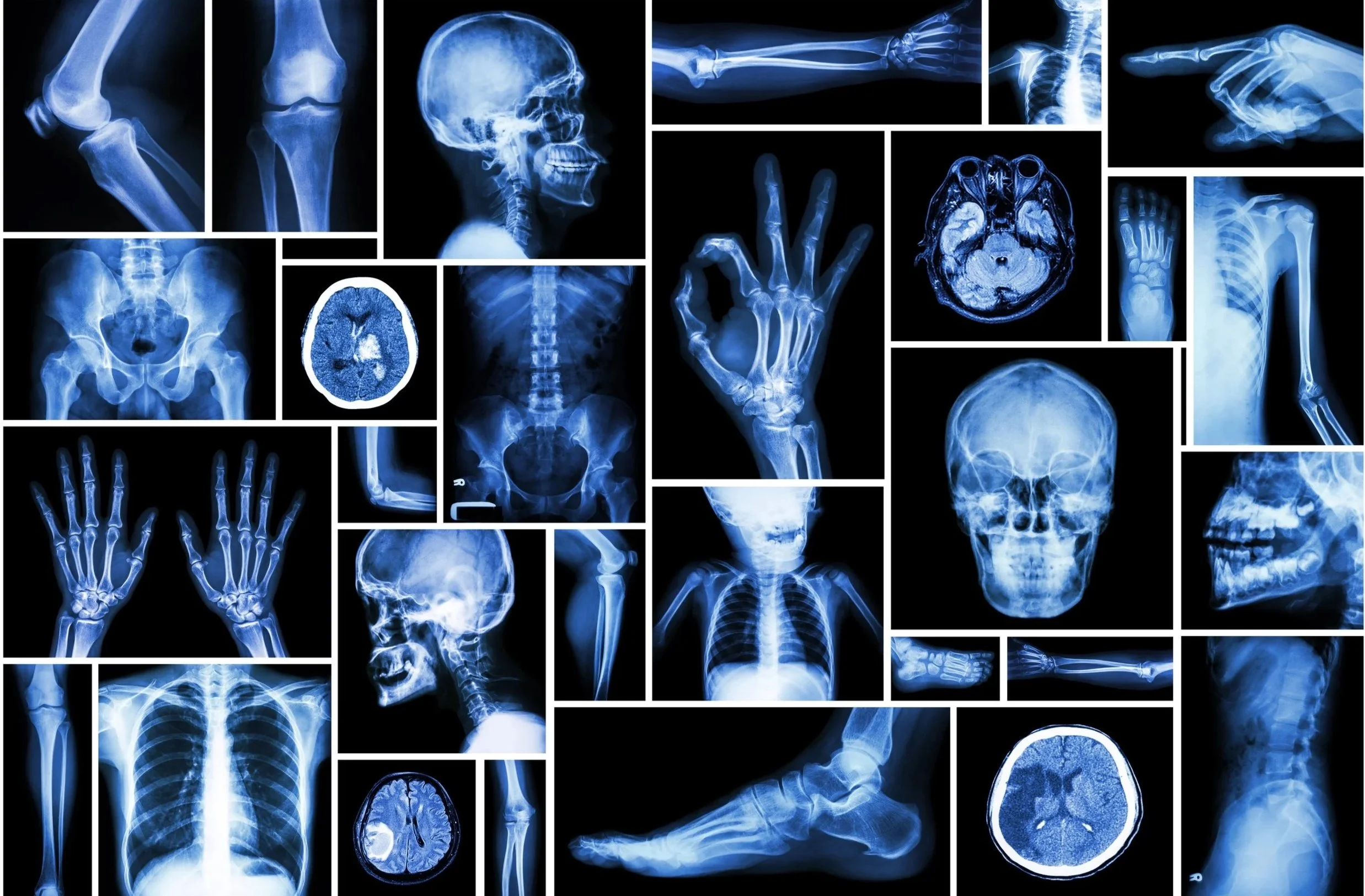
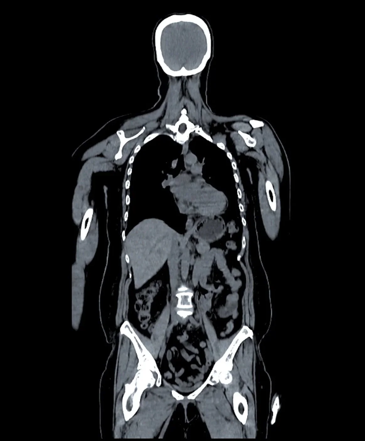
Medical Imaging Modalities
CT is an advanced imaging modality that uses X-rays and computer processing to create detailed cross-sectional images of the body, helping evaluate a wide range of conditions including trauma, infection, cancer, vascular disease, and other acute or chronic abnormalities.
Computed Tomography (CT)
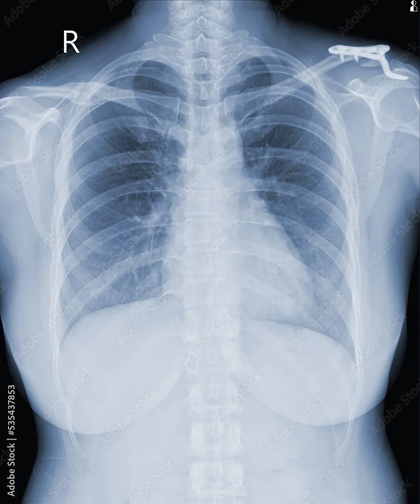
X-ray radiography is a foundational imaging modality that uses ionizing radiation to produce two-dimensional images of the body, commonly used to evaluate bones, the chest, and a wide range of acute and chronic conditions.
X-ray (Radiography)
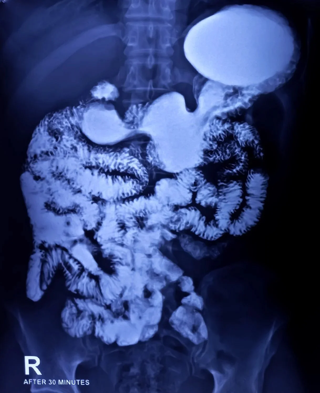
Fluoroscopy is an imaging modality that uses continuous or pulsed X-rays to produce real-time moving images, often used to evaluate swallowing, gastrointestinal function, joint or catheter procedures, and other dynamic processes within the body.
Fluoroscopy
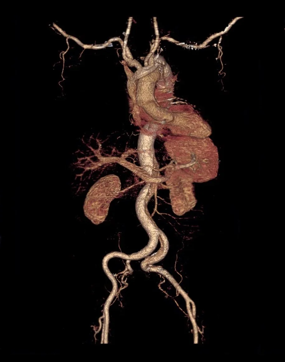
CTA is a contrast-enhanced CT study used to visualize blood vessels throughout the body and evaluate conditions such as aneurysm, dissection, stenosis, vascular occlusion, and other arterial or venous abnormalities.
Computed Tomography Angiography (CTA)
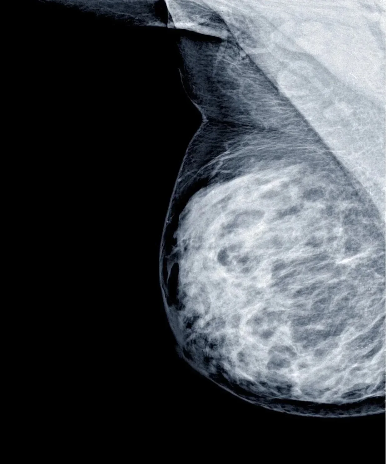
Mammography is a specialized X-ray imaging modality used to evaluate breast tissue for cancer and other abnormalities. It includes both standard 2D mammography and digital breast tomosynthesis (3D mammography), which acquires multiple low-dose images to improve lesion detection and breast tissue visualization.
Mammography including Tomosynthesis
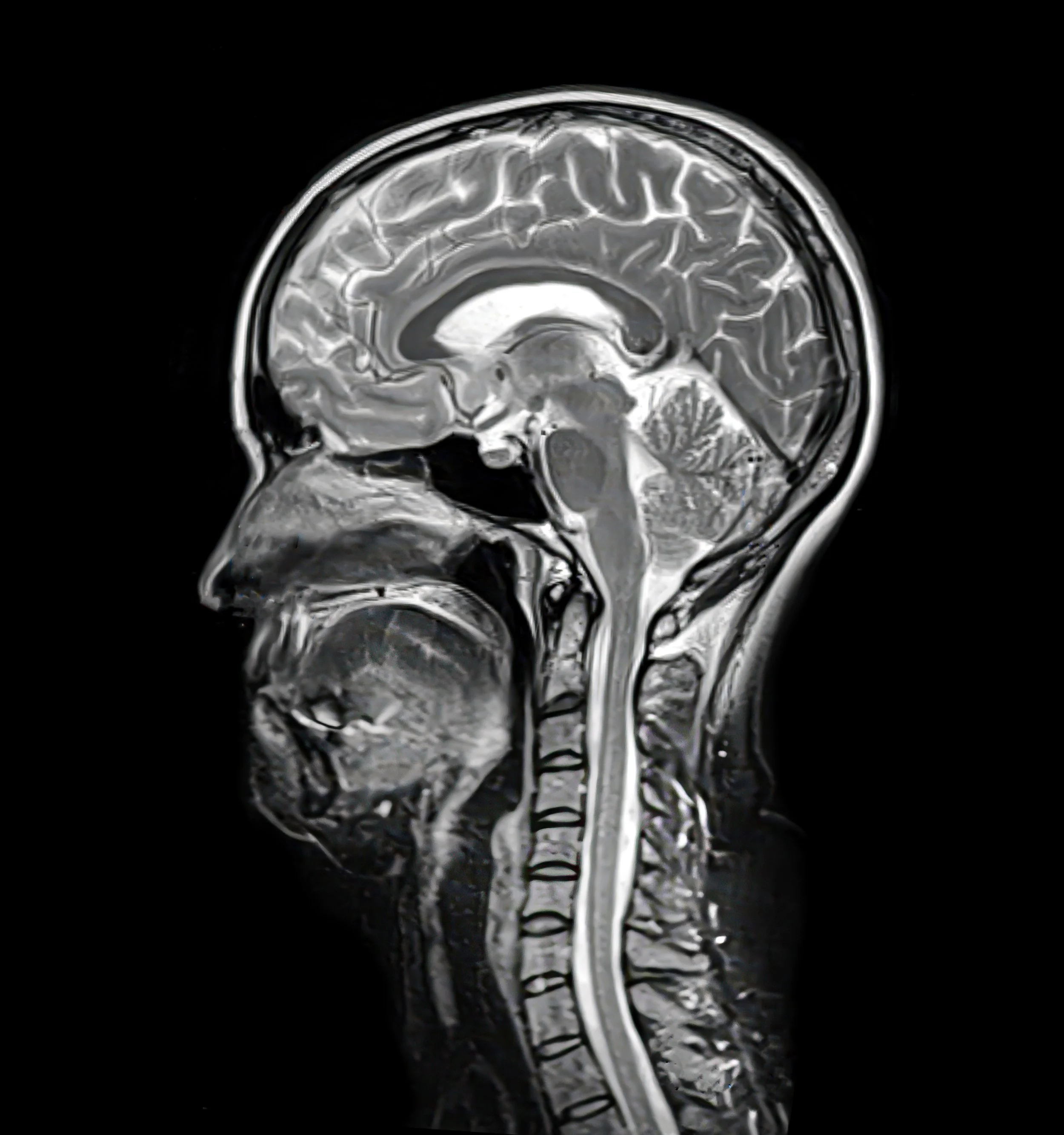
Magnetic Resonance Imaging (MRI) is an advanced imaging modality that uses powerful magnets and radiofrequency signals to produce detailed images of soft tissues, organs, the brain, spine, joints, and other internal structures.
Magnetic Resonance Imaging (MRI)
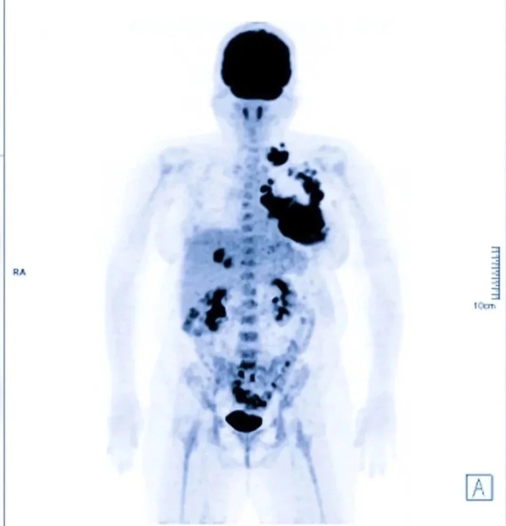
PET-CT is an imaging study that combines metabolic information from PET with anatomical detail from CT, allowing more precise evaluation of cancer, infection, inflammation, and other active disease processes.
Nuclear Medicine/ PET-CT Imaging
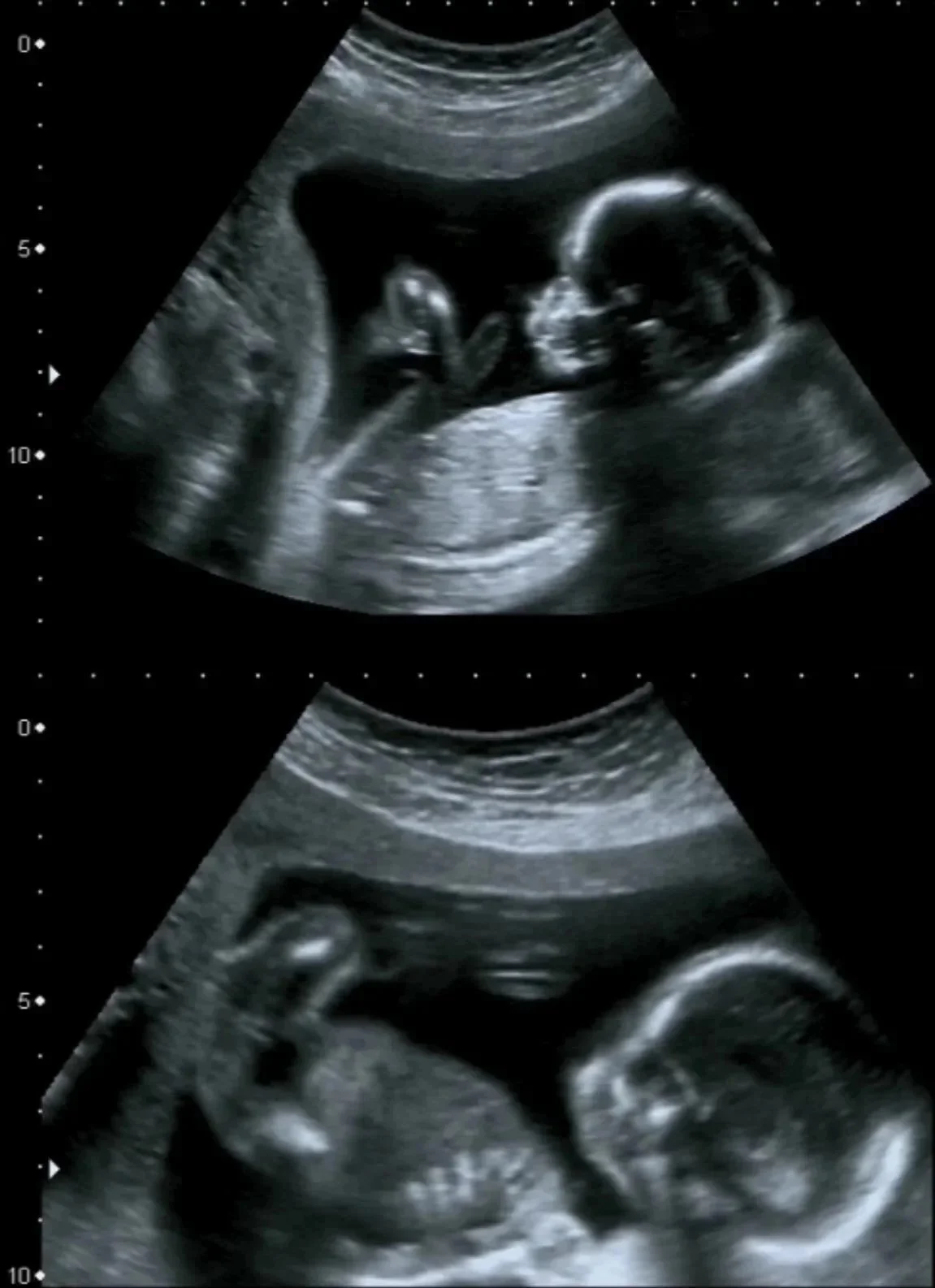
Ultrasound
Ultrasound is an imaging modality that uses sound waves to produce real-time images of organs, soft tissues, blood flow, and developing fetal structures. It includes gray-scale, Doppler, color Doppler, power Doppler, spectral Doppler, duplex ultrasound, and elastography.
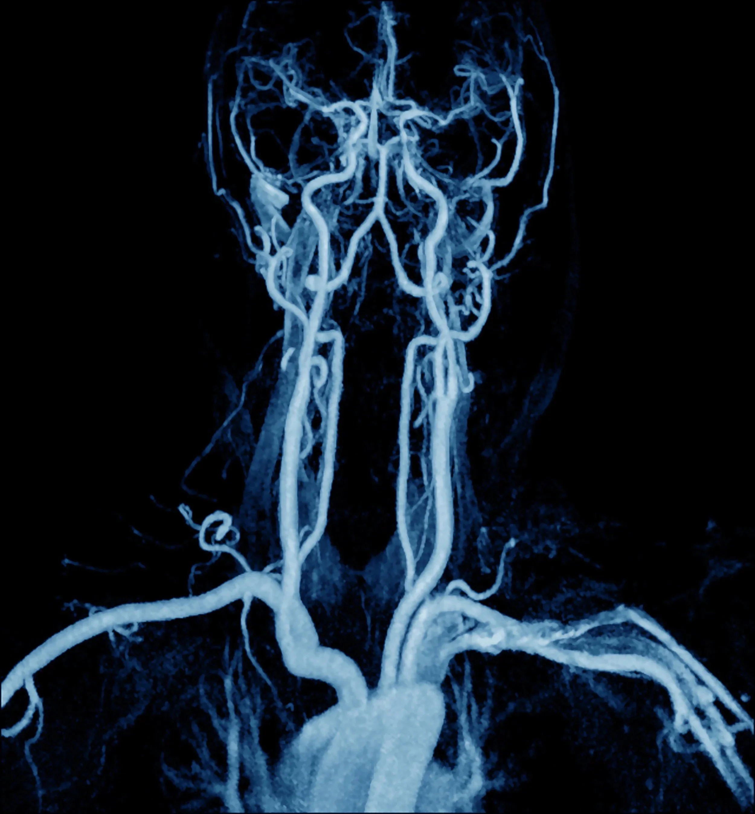
Magnetic Resonance Angiography (MRA) is an MRI-based technique used to visualize blood vessels and evaluate conditions such as aneurysm, stenosis, occlusion, dissection, and other vascular abnormalities.
Magnetic Resonance Angiography (MRA)
Contact
To inquire about radiology legal consulting services, please complete the form and provide a few details about your case. Dr. Milosavljevic will personally review your submission and respond promptly, typically within 48 hours.